
The Management of Open Chest Injury
16th December 2019 updated 18th March 2024
Traumatic Chest Injury accounts for 25% of all traumatic deaths (1) and pneumothorax is the single most common manifestation of intrathoracic blunt chest injury. (2)
An open pneumothorax allows air to enter during inspiration and exit during expiration causing the lung to collapse and fall away from the chest wall creating a pleural space. This free-flowing wound is often referred to as a “sucking chest wound”.
Two examples of open pneumothoraces - notice the small amount of blood which has a propensity to block vented chest seals if not closely monitored.
The larger the hole, the more likely that air will enter the thorax through the wound during inspiration rather than through the trachea and into the lungs. (3)
It is often stated that if the hole is equal to or greater than two-thirds the diameter of the trachea, air will preferentially enter the chest wall during inspiration, rather than the trachea, resulting in respiratory failure and eventual cardiopulmonary arrest. (4) The average human trachea is between 10 to 27 mm based on a study of 808 adults.(5) That would mean you seal a chest wound if it is 6 mm in diameter in some cases and other times not until it is 18 mm which is not a robust decision-making tool.
Open chest injuries should be managed based on the efficacy of the casualty’s breathing, not the size of the hole.
Tension Pneumothorax
A tension pneumothorax occurs when an injury to the lung, bronchi, or trachea allows a continuous leak of air into the pleural space and progressively collapses the lung when air cannot escape. A build-up of positive pressure within the plural space will further collapse the lung on the affected side to the point that anatomical structures are shunted away, compressing the otherwise unaffected lung and the heart, reducing cardiac output.
How this develops differs depending on whether the casualty is spontaneously breathing or being ventilated with a BVM. (6)
In spontaneously breathing casualties, the pathophysiology from the negative pressure of inspiration is gradual hypoxia from reduced cardiac output and structural collapse (7) progressing to respiratory failure and eventually respiratory arrest. (8,9)
In ventilated patients, the positive pressure forced into the pleural space from the damaged lung, bronchi or trachea can lead to a rapid development of tension pneumothorax with a rapid drop in SpO2 and is followed quickly by hypotension. (10)
Signs & Symptoms of Tension Pneumothorax
Early signs of a tension pneumothorax include:
Tachypnea (fast breathing)
Dyspnea (difficulty in breathing)
Tachycardia (fast pulse above 100pbm)
Hypoxia
This can develop into Becks Triad of
Distant heart sounds (only observable with a stethoscope and only practically observable in a quiet environment)
Distended neck veins (reduced cardiac output causes a build-up of returning blood in the superia vana cava that becomes evident as it backs up into the jugular veins)
Hypotension (low blood pressure – rare in the spontaneously breathing casualty (11))
Chest Seals
The simple solution would appear to be to seal the hole with an occlusive (air-tight) dressing. Although the use of an occlusive dressing is effective at sealing a pneumothorax, it is completely useless for prevention of a tension pneumothorax and, if the casualty is being ventilated with a BVM, an occlusive dressing can actually cause tension pneumothorax. (12,13)
There are a number of commercially available vented chest seals on the market. One of the first was the Asherman Chest Seal which relies on a flutter valve akin to a whoopee cushion which allows positive pressure to vent out of the plueral space but prevents airflow into the pleural space. One of the early criticisms of the design was that it just didn’t stick to sweaty, bloody or soiled skin making it effectively useless in a pre-hospital environment. The more rigid nature of plastic dressing meant that when folded in storage, any creases left in the dressing prevented a good seal being made which reduced its efficacy. An improvement was the Bolin Chest Seal which utilises plastic one-way valves, a more sticky adhesive and a more pliable material which did not crease.
In tests both of these designs were inferior in adhesion to soiled skin compared to more recent designs such as the Russell, Hyfin, SAM or FastBreathe*. (14)
Another issue with both the Asherman and Bolin Chest Seal were their inability to allow blood through the valves. Accumulated blood clogged the air passage and led either to development of tension hemopneumothorax, or more often, caused detachment of chest seals from skin and loss of function. (15)
More recent designs rely on two laminar layers which allow for one-way air flow but do not appear clog (and therefore become occlusive) or detach due to the introduction of blood. (12)
In tests both the Sentinel* and Russell received 100% success rates (n=6), 67% for Hyfin and 25% for the SAM. (16)
The Celox Foxseal shows promise but there is no comparative trails to date.
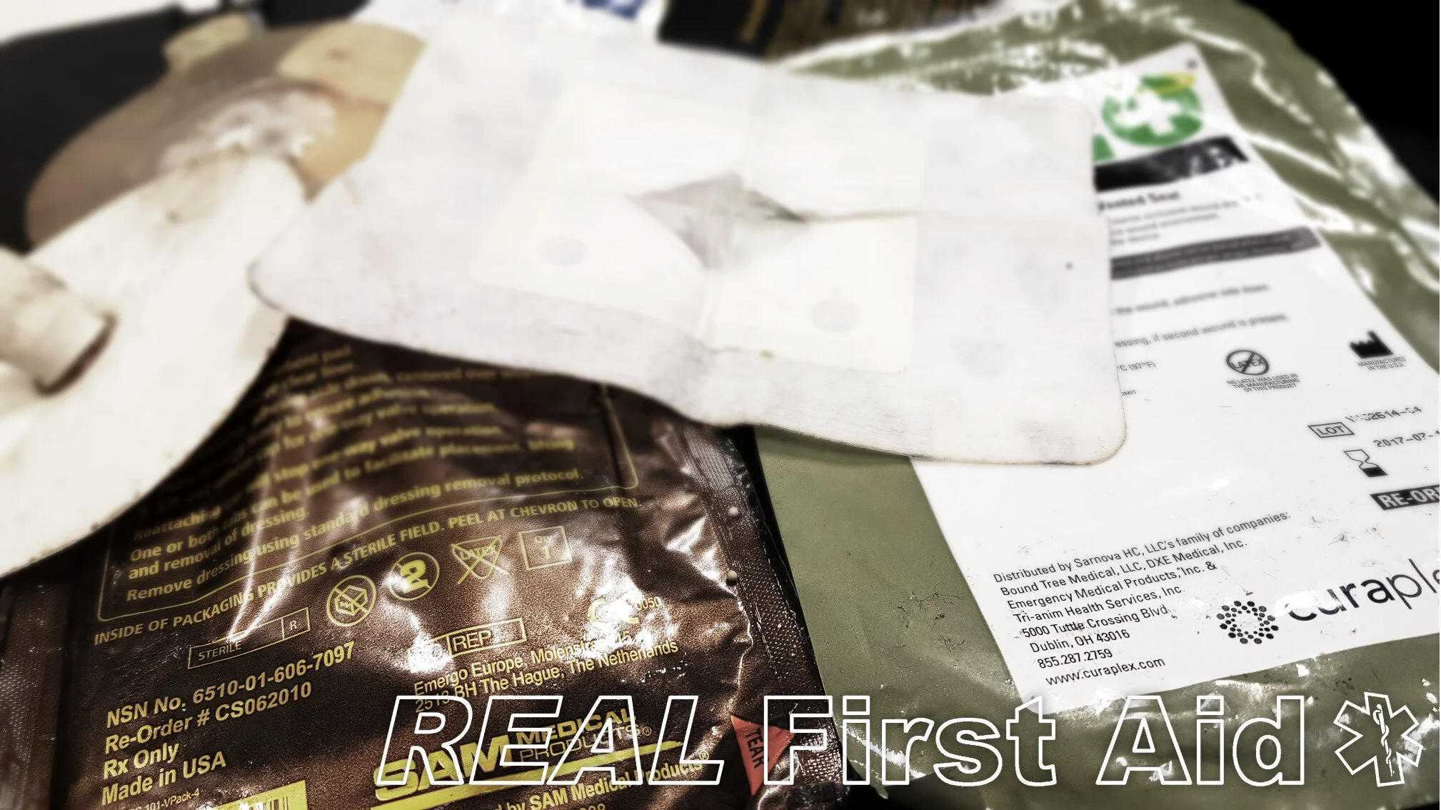
Vented Chest Seals - Outer Packaging L-R: SAM, Russell, Hyfin, HALO Vent, Bolin, Asherman.
Vented Chest Seals L-R: SAM, Russell, Hyfin, HALO Vent, Bolin, Asherman.
Improvised 3 sided versus Vented
Improvised 3 Sided dressings are not recommend. They are often difficult to adhere, ineffective and difficult to improvise in time-critical scenarios. (17-19)
An example of an imporvised chest seal which is failing to prevent air being sucked into the chest.
Treatment
The optimal position for gas exchange is sitting up, or lying with the healthy lung down (18, 20-23). This is unlikely to be possible in the case of patients who are hypovolaemic, in whom spinal fractures cannot be excluded, where lung injury has caused airway bleeding, or for practical reasons of safety during transfer.
Provide supplemental oxygen to ensure saturations remain at 94% or above.
The current 2021 ERC guidelines recommend open chest wounds should be “…exposed to freely communicate with the external environment” to prevent the development of a tension pneumothorax. (24)
Use a vested chest seal if one is available (6, 24), we recommend the Russel or Foxseal.
If a vented chest seal is not available – do not improvise (17-19)
If the casualty is breathing spontaneously or being ventilated, leave the chest injury open to prevent tension pneumothorax (24)
If the casualty is breathing spontaneously but is showing signs of respiratory distress, consider partially occluding the wound to allow reduced free-flowing movement of air to prevent tension pneumothorax. (6)
A vented chest seal is not a ‘fit and forget’ solution; as well as air passing through the vent will be blood and all vented chest seals have the potential to clog with coagulated blood and become occlusive, potentially causing a tension pneumothorax. If this occurs, remove the clogged chest seal and reapply to partially cover the hole.
Consider the chest as two separate boxes:
An open wound on each side should both be left open or covered with a vented chest seal.
If there are multiple open wounds on either side, leave one wound open (or use a vented chest seal) on each side. Completely seal all other holes.
The chest is the entire area above the diaphragm including the sides and the back. Check the whole chest for injuries and treat both sides accordingly (including the back).
*FastBreathe and Sentinel chest seals are not commonly available in the UK

References
Livingston DH, Hauser CJ. (2003) “Trauma to the chest wall and lung”. In: Moore EE, Feliciano DV, Mattox KI, eds. Trauma. 5th ed. New York, NY: McGraw-Hill Medical. 507–537.
Brasel KJ, Stafford RE, Weigelt JA, Tenquist JE, Borgstrom DC. (1994) “Treatment of occult pneumothoraces from blunt trauma”. Journal of Trauma. 46:987–991.
Hodgetts T, Hanlan C, Newey CI. (1999) “Battlefield First Aid: A simple, systematic approach for every soldier”. Journal of the Royal Army Medical Corps. 145:55-59.
American College of Surgeons Committee on Trauma. Thoracic trauma. In: ATLS: advanced trauma life support for doctors: student course manual. 8th ed. Chicago: American College of Surgeons Committee on Trauma; 2008:87
Breatnach E, Abbott GC, Fraser RG. (1984) “Dimensions of the normal human trachea”. American Journal of Roentgenology. May;142(5):903-6.
Littlejohn, LF. (2017) “Treatment of Thoracic Trauma: Lessons From the Battlefield Adapted to All Austere Environments”. Wilderness & Environmental Medicine. 28, S69–S73
Carvalho P, Hilderbrandt J, Charan NB. (1996) “Changes in bronchial and pulmonary arterial blood flow with progressive tension pneumothorax”. Journal of Applied Physiology. 81:1664–1669.
Rutherford B, Hurt HH Jr, Brickman RD, Tubb JM. (1968) “The pathophysiology of progressive, tension pneumothorax”. Journal of Trauma. 8:212–227.
Gustman P, Yerger L, Wanner A. (1983) “Immediate cardiovascular effects of tension pneumothorax”. American Review of Respiratory Disease. 127:171–174.
Coats TJ, Wilson AW, Xeropotamous N. (1995) “Pre-hospital management of patients with severe thoracic injury. Injury. 2:581–585.
Barton ED. (1995) “Tension pneumothorax”. Current Opinion in Pulmonary Medicine. 5:269–274.
Kotora, Joseph G. et al. (2013) “Vented Chest Seals for Prevention of Tension Pneumothorax in a Communicating Pneumothorax“. Journal of Emergency Medicine. 45(5);686-694
Kheirabadi BS, Terrazas IB, Koller A, Allen PB, Klemcke HG, Convertino VA, Dubick MA, Gerhardt RT, Blackbourne LH. (2013) “Vented versus unvented chest seals for treatment of pneumothorax and prevention of tension pneumothorax in a swine model”. Journal of Trauma and Acute Care Surgery. Jul;75(1):150-6
Arnaud F, Tomori T, Teranishi K, Yun J, McCarron R, Mahon R. (2008) “Evaluation of chest seal performance in a swine model: comparison of Asherman vs. Bolin seal. Injury. 39(9):1082-8
Arnaud F, Jeronimo-Maudin E, Higgins A, Kheirabadi B, McCarron R, Kennedy D, Housler G. (2016) “Adherence evaluation of vented chest seals in a swine skin model”. Injury. 47(10):2097-2104
Kheirabadi BS, Terrazas IB, Miranda N, et al (2017) “Do vented chest seals differ in efficacy? An experimental evaluation using a swine hemopneumothorax model”. Journal of Trauma and Acute Care Surgery. 83(1):182-189
BET 3: “In a penetrating chest wound is a three-sided dressing or a one-way chest seal better at preventing respiratory complications?” Emergency Medicine Journal 2012;29:342-343.
Lee C, Revell M, PorterK, et al. (2007) “The prehospital management of chest injuries: a, consensus statement”. Faculty of Pre-hospital Care, Royal College of Surgeons of Edinburgh. Emergency Medical Journal. 24:220–4.
Butler FK, Dubose JJ, Otten EJ, et al. (2013) “Management of Open Pneumothorax in Tactical Combat Casualty Care”. TCCC Guidelines Change 13-02. Journal Special Operations Medicine. 13(3):8186.
Dean E. (1985) “Effect of body position on pulmonary function”. Physical Therapy. 65(5):613-618.
Clauss RH, Scalabrini BY, Ray JF, et al. (1968). “Effects of changing body position upon improved ventilation- perfusion relationships”. Circulation. 37(Suppl2):214-217.
Remolina C, Khan AV, Santiago TV, et al. (1981). “Positional hypoxemia in unilateral lung disease”. New England Journal of Medicine. 304:523-525.
Sonnenblick M, Melzer E, Rosin AJ. (1983) “Body positional effect on gas exchange in unilateral pleural effusion”. Chest. 83:784-786.
Zideman DA, Singletary EM, Borra V, Cassan P, Cimpoesu CD, De Buck E, Djarv T, AJ Handley, Klaassen B, Meyran D, Oliver E, Poole K. (2021) “European Resuscitation Council Guidelines for Resuscitation 2021 Section 9. First aid”. Resuscitation. 161. p274